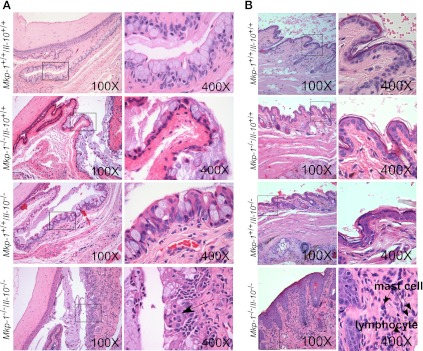
Fig. 6.
Double knockout mice exhibit conjunctival and eyelid lesions. Mkp-1+/+/Il-10+/+, Mkp-1−/−/Il-10+/+, Mkp-1+/+/Il-10−/−, and Mkp-1−/−/Il-10−/− mice were housed in a SPF environment and were euthanized at 3 mo of age. Eyes and periocular tissues were excised, routinely fixed, and embedded, and sections were stained with H&E. A: conjunctival lesions in double knockout mice. Mkp-1+/+/Il-10+/+ mice lacked conjunctival lesions (top row). Mkp-1−/−/Il-10+/+ and Mkp-1+/+/Il-10−/− mice showed moderately increased goblet cell numbers and mild increased mucus accumulation in the conjunctival space (2nd and 3rd rows). The conjunctival mucosa of Mkp-1−/−/Il-10−/− mice was markedly thickened with moderate goblet cell hyperplasia and moderate to marked mucus accumulation in the conjunctival space (bottom row). Additionally, the conjunctival stroma was expanded by mild edema and infiltrated by neutrophils, with fewer lymphocytes and plasma cells. B: eyelid lesions in Mkp-1/Il-10 double knockout mice. The haired skin of double knockout mouse eyelids mice was markedly thickened and infiltrated by lymphocytes and plasma cells, with scattered clusters of mast cells. Wild-type, Mkp-1−/−/Il-10+/+, or Mkp-1+/+/Il-10−/− mouse eyelids lacked lesions. Column on the right contains high-resolution images of the boxed areas in left column.

