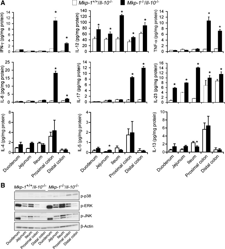
Fig. 7.
Colons from Mkp-1/Il-10 double knockout mice contain higher levels of proinflammatory cytokines. The intestine of both double knockout and Il-10 knockout mice were divided into duodenum, jejunum, and ileum, as well as proximal and distal colons. Tissues were homogenized to extract soluble proteins. A: levels of cytokines in distinct intestinal regions. IFN-γ, IL-4, IL-5, IL-6, IL-12 (p70), IL-13, IL-17, IL-23 (p19/p40), and TNF-α in the tissue extracts were assessed by ELISA. Values were normalized to total protein content in the tissue homogenates and presented as means ± SE from at least 3 different animals. *P < 0.05, compared with level in the Mkp-1+/+/Il-10−/− tissue (Student's t-test). B: activities of distinct MAPKs. The active forms of MAPKs in the tissue homogenates were detected by Western blot analyses, using antibodies against phospho-MAPKs. Comparable protein loading was verified by Western blot analysis using an antibody against β-actin (bottom). Presented are the representative results of at least 3 experiments.

