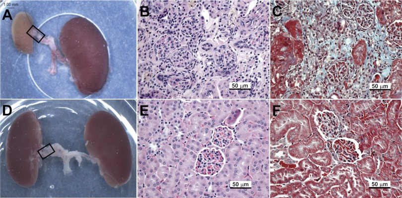
Fig. 6.
The cuffed (stenotic) kidney of Smad3 KO mice does not develop atrophy, despite reduced blood flow. Representative images of gross and microscopic changes in WT (A–C) and Smad3 KO (D–F) kidneys 6 wk after RAS cuff surgery are shown. Gross images (A and D) were captured at time of death; RAS tubing cuff is outlined by a black rectangle. Histological sections were stained with hematoxylin and eosin (B and E) or Masson's Trichrome (C and F).

