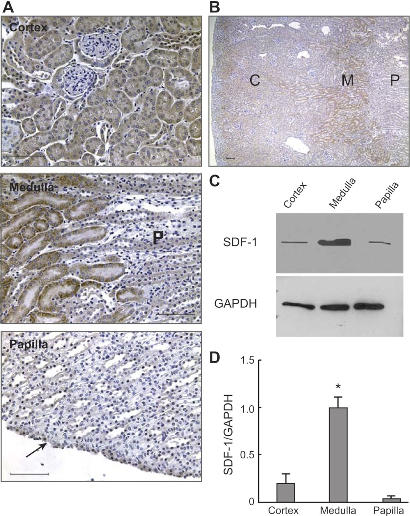
Fig. 7.
Regional location of SDF-1 in the adult kidney. A: Immunohistochemistry for SDF-1 in the cortex (top), medulla (middle), and papilla (bottom). As shown, SDF-1 was widely distributed in the cortex and medulla, whereas little signal was detected in the papilla (except in the uroepithelium; arrow). Note in the middle picture that the strong signal for SDF-1 in the medulla (left) sharply disappears in the top papilla, marked P. Bars = 50 μm. B: the greater prominence of the SDF-1 signal in the medulla than in other parts of the kidney is illustrated in this low-power image, which shows, from the left, the cortex (C), medulla (M), and papilla (P) regions of the same kidney. Kidney sections incubated with identical amounts of irrelevant rabbit IgG and simultaneously processed with those with the SDF-antibody gave no signal (not shown). Bar = 200 μm. C: representative immunoblot for SDF-1 and GAPDH in different regions of the kidney. D: relative abundance of SDF-1 vs. GAPDH in different regions of the kidney showed that the medulla had a significantly greater amount of SDF-1 than both the cortex and the papilla (*P < 0.01). Data derive from 4 individual blots, each from an independent rat. In each blot, protein homogenates from the 3 different regions of the kidneys were simultaneously analyzed. Each lane for SDF-1 contained 50 μg of protein homogenate. Because of the large signal obtained with the antibody to GAPDH, for proper quantification of this protein's abundance in the sample, only one-fifth (i.e., 10 μg) of the protein homogenates were loaded on the gels.

