
Fig. 4.
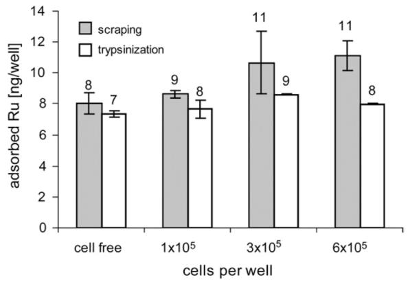
Influence of different degrees of cell confluence on adsorption of KP1019 to the plates. No cells, 1 × 10⁵, 3 × 10⁵ or 6 × 10⁵ cells were seeded and exposed to 50 μM of KP1019 for 2 h. Cells were scraped off or trypsinized after exposure, and wells were subsequently incubated with nitric acid in order to desorb ruthenium from the plates.