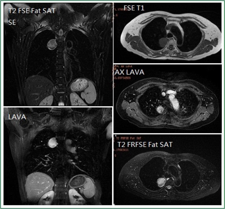
Figure 2.
Chest MRI findings of a middle-aged female patient. A round-like lesion with long T1 and T2 signals at the upper posterior portion of the right mediastinum (near the third thoracic vertebra) is observed. The lesion has a size of 2.9 cm × 3.1 cm × 3.5 cm, clearly defined margin, and inhomogeneous internal signal intensity. The lesion shows slightly high signal intensity on DWI. Dynamic enhancement imaging shows that the lesion is slightly enhanced in the arterial phase, and such enhancement becomes even more obvious in the portal venous phase and delayed phase; meanwhile, inhomogeneous internal enhancement is also observed.

