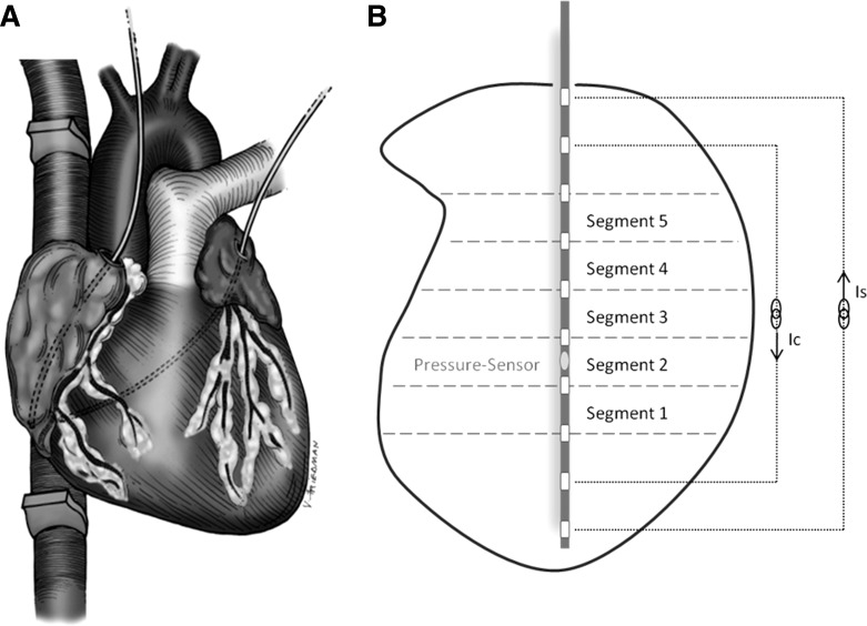
Fig. 2.
A: intraoperative setting: conductance catheters were placed in the right and left atrium with the tips resting at the inferior caval-atrial junction and the orifice of the right inferior pulmonary vein, respectively. Ultrasonic flow probes were placed around the superior and inferior vena cava. B: total atrial volume was divided into 5 segments. Conductance was measured using dual-field technology. Ic/Is, current yielding dual field tachnology.

