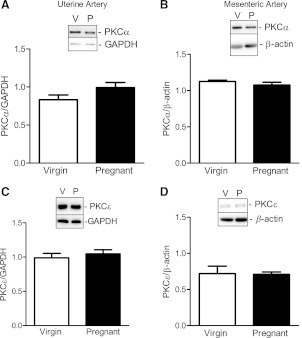
Fig. 5.
Pregnancy does not change the expression of PKC-α and PKC-ε in nonstimulated UtAs and MAs. A–D: representative Western blots (insets) and corresponding densitometric analysis showing the expression of uterine (A) and mesenteric (B) PKC-α (82 kDa) and uterine (C) and mesenteric (D) PKC-ε (90 kDa) in arteries from virgin and pregnant rats. Results from uterine tissue were normalized to GAPDH (37 kDa), and results from mesenteric tissue were normalized to β-actin (42 kDa). Densitometric data are expressed as means ± SE; n = 4–7.

