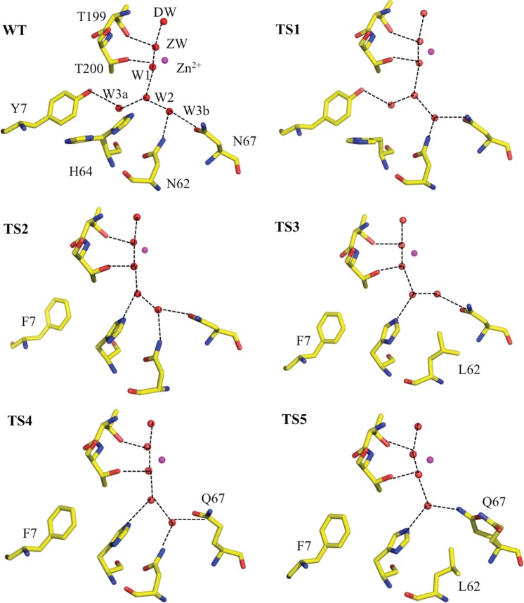
Fig. 2.
Ball-and-stick diagrams of wild type and stabilized variants of HCA II active sites. The Zn is shown as a magenta sphere and all the hydrophilic active site residues (and their counterparts in the mutants) are shown as sticks, waters are shown as red spheres. Residues and waters are as labeled and inferred H-bonds are shown as black dashed lines. Maps are omitted for clarity. Figures were generated and rendered with PyMOL (DeLano, 2002).

