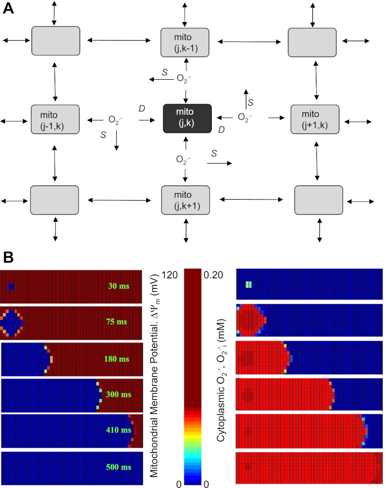
Fig. 1.
A: scheme of the two-dimensional (2-D) reaction-diffusion reactive oxygen species (ROS)-induced ROS release (RD-RIRR) mitochondrial network model. In the RD-RIRR model, neighboring mitochondria are chemically coupled with each other through superoxide anion (O2·−) diffusion. Light and dark gray indicate polarized and depolarized mitochondria, respectively. Arrows indicate release of superoxide anion (O2·−) and its effect on mitochondrial neighbors. D stands for extramitochondrial O2·− diffusion, and S for O2·− scavenging by Cu/ZnSOD and catalase. B: simulation of spatial propagations of mitochondrial inner membrane potential (ΔΨm) depolarization wave (left) and ROS wave (right) by the 2-D RD-RIRR model. This network consisted of 500 mitochondria (10 × 50), in which 6 were initially depolarized, whereas the others were polarized. Figure was obtained from (98) with permission.

