
Figure 1. Developmental progression of myogenesis and myogenic gene expression.
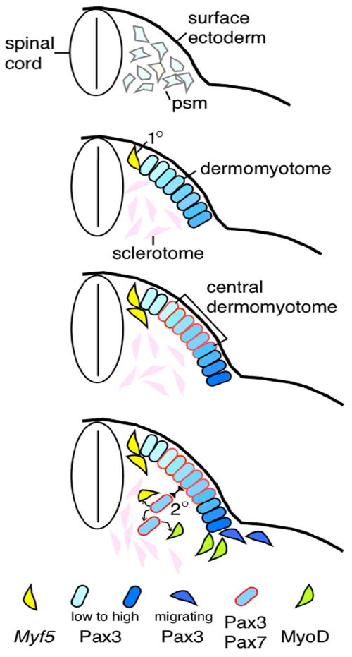
Top panel: presomitic mesoderm cells express Pax3 (pale blue) and low levels of Myf5 (pale yellow) transcripts. Epithelial somite stage is omitted. Second panel: appearance of sclerotome, dermomyotome (Pax3+), and primary (1°) myogenic cells (Myf5+) at the dorsal medial edge. Pax3 expression is at a higher level at the lateral edge. Third panel: Pax7 expression emerges and overlaps with Pax3+ cells in the central dermomyotome in more mature somites. Bottom panel: Vertical division of the central dermomyotomal cells, which give rise to the secondary (2°) myogenic progenitors. These cells are presumed to give rise to more progenitors and Myf5+ or MyoD+ myogenic cells. The lateral Pax3+ cells give rise to migrating myoblasts entering the ventral body wall and limbs. Keys to cells with specific gene expression are at the bottom.