
Figure 2. Pax7 expression is detected in satellite cells of the mouse tibialis anterior muscle.
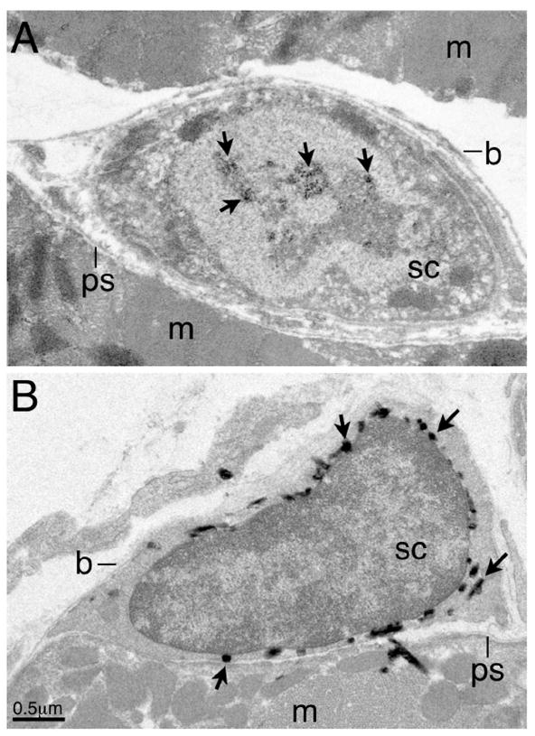
(A) Immuno-EM detection of endogenous Pax7 by a monoclonal antibody (DSHB), followed by a HRP-conjugated goat anti-mouse IgG1 antibody (Molecular Probes) and enzymatic reaction using the DAB substrate (Vecta Lab). The sample was fixed in Zamboni’s fixative following the protocol in Chen et al. (2007). (B) TEM of samples from tamoxifen-treated Pax7-Cre-ERT2;LacZ reporter mice, fixed in 4% paraformaldehyde, reacted with X-gal substrate, following the procedure in Kanisick et al. (2009). Abbreviations: m, muscle fiber, b, basement membrane; ps, plasmalemmal surface; sc, satellite cell. Arrows in (A) indicate reacted DAB deposits in the nucleus, in (B), reacted X-gal precipitates in the cytoplasm. Some X-gal precipitates are often seen next to satellite cells, likely due to substrate diffusion during enzymatic reaction. Scale bar in (B).