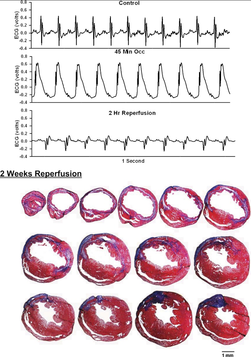
Fig. 4.
ECG before, during, and after occlusion of the LAD in a chronically instrumented, intact, conscious, unrestrained mouse (top) and photomicrographs of histological sections taken from the infarcted heart 2 wk postocclusion (bottom). See Fig. 3 legend for more details.

