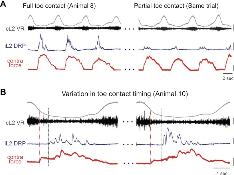
Fig. 8.
Role of contralateral toe contact in DRP generation. A: L2 DRP relative to ipsilateral ventral root during locomotion with full contralateral toe contact (MTP to toe tip, left); 130 s later in the same locomotor bout (right), the toe moved to the front of the force platform such that only a small portion of the toe contacted the plate. The reduction in toe contact, and likely toe afferent input, resulted in reduced DRPs. Scale bars are 400 μV, 5 mN, and 2 s. B: in another animal, toe contact occurred after force onset due to mid-foot contact. Two examples of this are shown. Black dashed vertical lines indicate toe contact time. The DRP occurred immediately after the toe contacted the plate rather than at force onset as indicated by red dashed line. Scale bars are 200 μV, 2 mN, and 1 s.

