
Figure 3. HIF-1a independent upregulation of VEGF expression in the aged rat cortex during chronic hypoxic exposure.
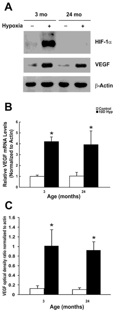
A, Representative Western blot analysis of HIF-1α, VEGF, & β-actin following 10 days of hypoxic exposure in the young and old F344 rats. Data are representative of at least 3 experiments. B, HIF-1 independent transcriptional activation of vegf in the aged rat cortex during chronic hypoxic exposure. Results were expressed as fold induction compared with cortical samples of brains exposed to normoxia and normalized to β-actin mRNA. Each individual value represents the mean +/− SD from three independent experiments from at least three different rats. *p < 0.05 compared with normoxia. C, Optical density ratios of VEGF normalized to β-actin before and after hypoxic exposure as a function of age. Each value represents the mean +/− SD from at least three rats. *p < 0.05 compared with control (non-hypoxic) cortical samples.