Fig. 1.
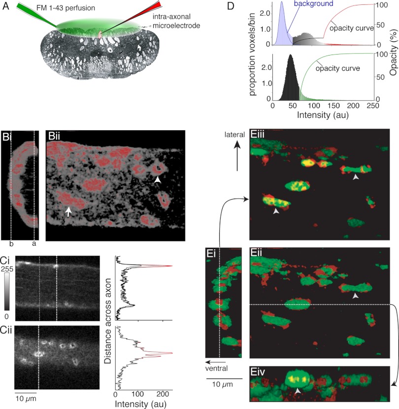
Distribution of phalloidin labeling and FM-labeled vesicle clusters in axons. A: recording schematic. FM1-43 was superfused over the ventral spinal cord (green). Axons impaled with microelectrodes containing KCl (3 M) were loaded with FM1-43 by intracellular stimulation, and the tissue was cleared of dye with Advasep-7. Axons were reimpaled (electrode contained Alexa Fluor 568 phalloidin; red) for injection into the axon. Confocal z-sections were taken of live axons. B: phalloidin labels both synaptic structures and cortical actin. Bi: 3-dimensional (3D) reconstruction viewed along length of the axon (caudal-rostral) showing ventral half of axon and phalloidin labeling (ventral-left). Red, intense labeling at puncta; gray, dimmer, diffuse labeling under plasma membrane—cortical actin [see D for color look-up table (LUT)]. Bii: view of same axon from the spinal ventral surface. Ci: optical section from dashed line a in Bi. Graph shows intensity profile through the section at dashed lines [white, through active zone (graph color from same LUT as B); gray, outside synaptic regions]. This graph is in gray. Cii: optical section b in Bi. Graph of intensity profile along dashed line in image. Colors from LUT as for Ci. D, top: voxel intensity histogram from data set in B. Background noise sampled from outside axons (blue line). This matches background noise under the image (light blue bars). Other histogram bar colors represent the LUT used to generate 3D reconstructions. Black to red curve represents opacity values used in B. Bottom: voxel intensity histogram for FM1-43 labeling; opacity curve to display FM1-43 in E in green. au, Arbitrary units. Ei: 3D overlay of phalloidin structures (red) and FM1-43 labeling (green) (line, section in Eiii). Eii: axon from spinal ventral surface (view from left of Ei) (line, position of section in Eiv). Eiii: view as Ei but top sections removed (cut at dashed line in Ei) to reveal phalloidin and FM1-43 colocalization (arrows: phalloidin, red; FM-loaded vesicles, bright green) at the cut at dashed line in Ei. Yellow, colocalization at cut. Eiv: sagittal cut of FM1-43 and phalloidin seen from lateral spinal cord with lower half below dashed line in Eii removed. Vesicle clusters are associated with phalloidin, sometimes spanning more than 1 phalloidin structure (arrowheads in Eii, Eiii). FM/phalloidin colocalized (yellow) can be contained within an FM1-43 puncta (arrowhead in Eiv).

