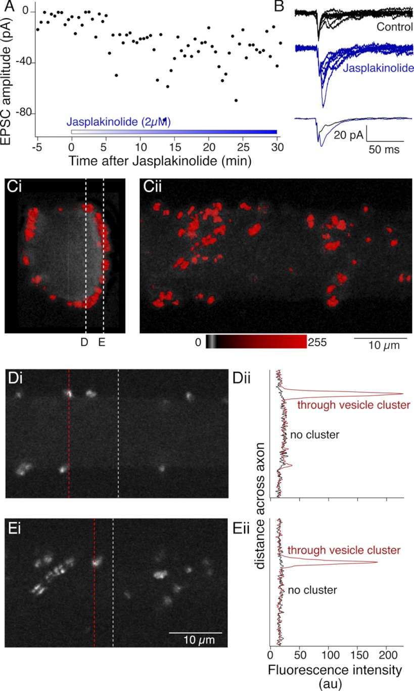
Fig. 10.
Jasplakinolide enhances synaptic transmission and prevents phalloidin labeling of cortical actin. A: paired cell recordings between reticulospinal axons and their whole cell clamped postsynaptic target neurons. Control responses were recorded to single presynaptic action potentials at 30-s intervals for 5 min. Jasplakinolide (2 μM) was added to the superfusate. Graph shows single EPSC peak amplitudes against time after addition of jasplakinolide. B: examples of 6 sequential EPSCs from control (black) and in jasplakinolide after 20 min of application (blue). Overlaid traces at bottom, averages of the EPSCs shown at top. C: after treatment with jasplakinolide (2 μM) for a minimum of 20 min, phalloidin was microinjected into axons through the recording pipette. Fluorescence was imaged confocally to reconstruct labeling in 3D 40 min after injection. Vesicle cluster-associated phalloidin was readily labeled. However, no cortical actin was seen. Ci: view along the length of the axon. Cii: view from ventral spinal cord. D and E. single optical sections from positions indicated by the dashed lines in Ci through the axon (Di) and at the ventral surface (Ei). Intensity profiles (Dii, Eii) are shown through these optical sections at the dashed lines in Di and Ei including clusters (red) and regions with no clusters (black). No cortical actin signal is seen at the axon membrane.

