Fig. 4.
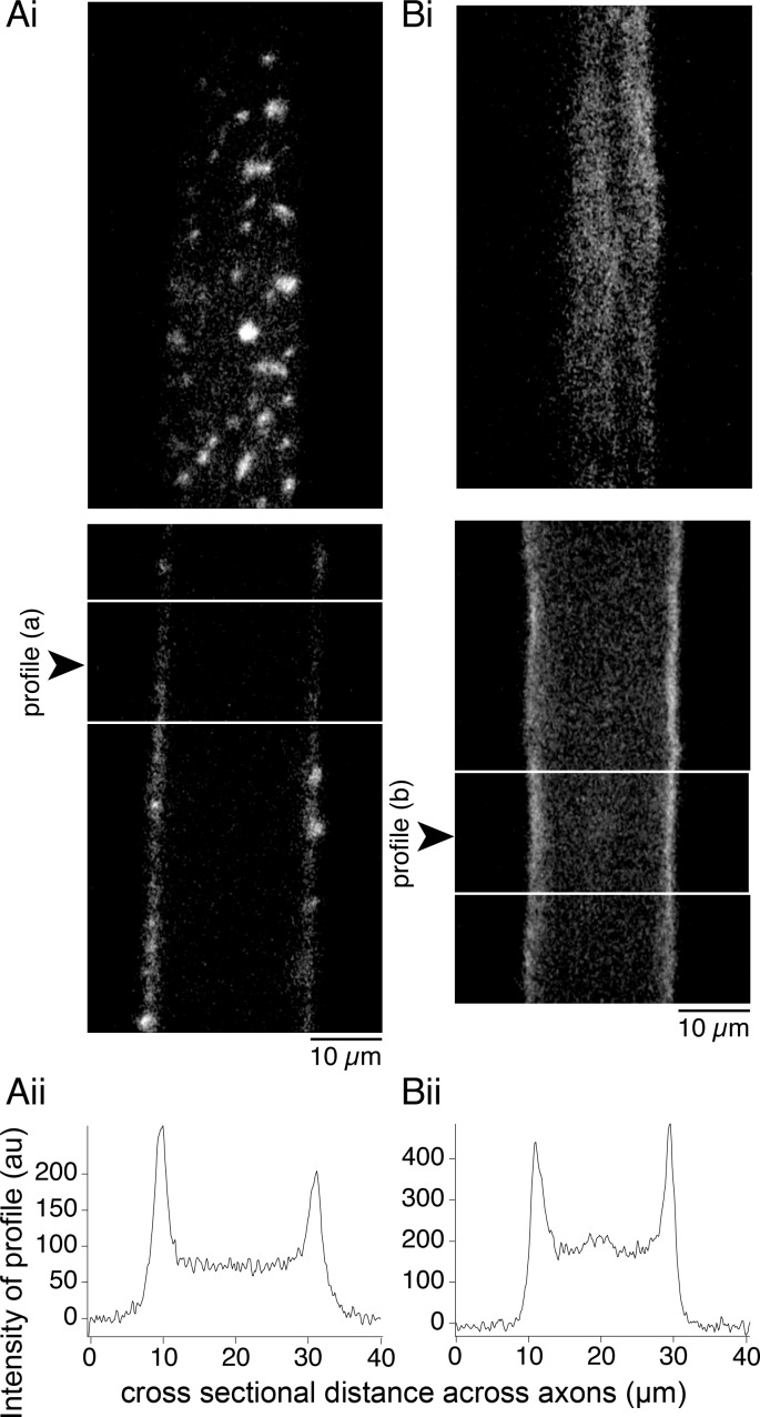
Latrunculin-A prevents incorporation of phalloidin at presynaptic terminals but not the cortical actin. Axons were recorded from with a microelectrode and injected with phalloidin (Alexa Fluor 488). Ai: control axon injected with phalloidin reveals vesicle cluster-associated fluorescent structures. Top: optical section through the ventral axon surface. Bottom: section through the center of the same axon. Aii: to quantify this, a profile of fluorescence intensity was measured across the longitudinal sections from between the horizontal white lines in Ai. Profile was placed to avoid presynaptic puncta. A fluorescence peak is seen at the plasma membrane. Bi: axon injected with phalloidin after latrunculin-A treatment (30 min, 12 μM) displays fluorescence with no clustering of phalloidin. Top: optical section at the ventral surface. Bottom: section through the center. Although no presynaptic structures are seen, plasma membrane fluorescence is visible. Bii: profile of fluorescence intensity from between lines in phalloidin-labeled axon (Bi) after pretreatment with latrunculin-A. Fluorescence peaks are seen at the membrane.

