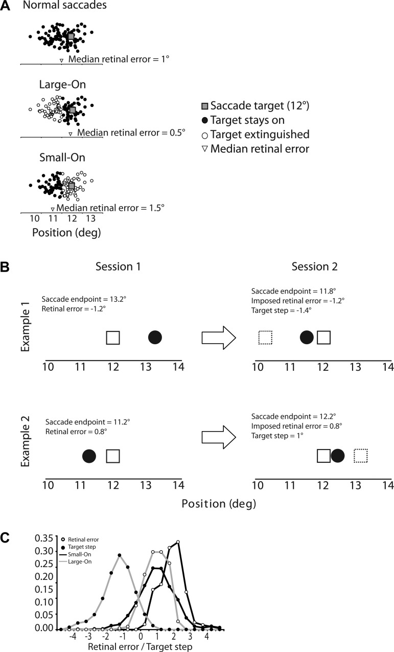
Fig. 1.
A: schematic illustration of amplitude-dependent visual feedback. In normal saccades (top), endpoints tend to undershoot the visual target. Thus the median retinal error tends to be positive. In the “Large-On” condition (middle), the visual target was available after the primary saccade only when its amplitude was larger than the median. Thus the distribution of retinal errors experienced by the subject was smaller than usual. In the “Small-On” condition (bottom), the visual target was available after the primary saccade only when its amplitude was smaller than the median. The distribution of retinal errors experienced by the subject was larger than usual. B: 2 examples of corresponding trials during the 2 sessions. The saccade target (open square) was always at 12°. In the first example (top), in session 1 the saccade endpoint (black dot) on trial n was 13.2°, which corresponds to a retinal error of −1.2°. In session 2, the saccade endpoint on trial n was 11.8°. To obtain a retinal error of −1.2°, the target had to be stepped back to position 10.6° (dashed square), a step of −1.4°. In the second example (bottom), in session 1 the saccade endpoint was 11.2° and retinal error was 0.8°. In session 2, since the endpoint was 12.2°, obtaining a retinal error of 0.8° required stepping the target forward by 1°. C: distribution of retinal errors and target steps in Large-On and Small-On conditions, pooled over all subjects. The zero mark on the x-axis corresponds to the target location; thus a step of 0 means that the target did not step, and a retinal error of 0 means that the saccade endpoint was at the same location as the postsaccadic target.

