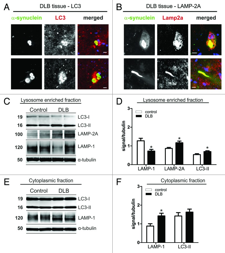
Figure 1. Immunohistochemistry for LC3 (A) and LAMP-2A (B) in α-synuclein positive Lewy bodies (LBs) in midbrain of DLB patients. Co-staining for both ALP markers was found in LBs. In addition, LAMP-2A was found in Lewy neurites (B, lower panel). Scale bars: 10 µm. Protein levels of LAMP-1, LAMP-2A and LC3 in cell lysates (lysosome enriched fraction, C and D; cytosolic fraction, E and F) of temporal cortex tissue of DLB cases and controls. Western blot analysis of ALP marker is shown for two representative DLB cases and controls (C and E). Quantitative densitometry revealed increased levels of LC3-II and LAMP-2A in the lysosome enriched fraction (D; DLB n = 12, controls n = 9), but not in the post-nuclear fraction (F; DLB n = 15, controls n = 11; D, upper panel). A LAMP-2A specific signal could only be detected in the lysosome enriched fraction. LAMP-1 was reduced in the lysosome-enriched fraction and increased in the post-nuclear fraction. (Student’s t-test, *p < 0.05).

An official website of the United States government
Here's how you know
Official websites use .gov
A
.gov website belongs to an official
government organization in the United States.
Secure .gov websites use HTTPS
A lock (
) or https:// means you've safely
connected to the .gov website. Share sensitive
information only on official, secure websites.
