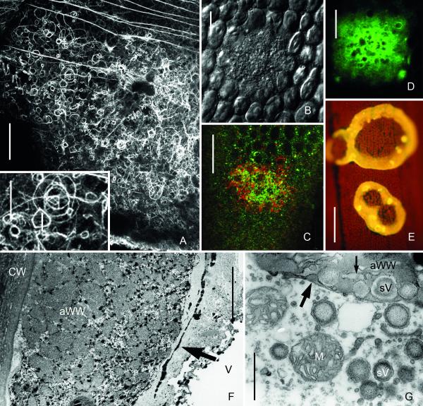
Fig. 4.
Amorphous wound walls induced by treatment of internodal cells of Nitella flexilis with 10−4 M chlortetracycline for one hour (A, F and G) and by 2 min local irradiation with UV of internodal cells of Chara corallina (B-E). (A) Actin cytoskeleton beneath a forming amorphous wound wall visualized by immunofluorescence. Note numerous actin rings and polygons (inset). Continuous actin bundles are present outside the wound. (B-D) Accumulation of secretory vesicles (B) and of FM1-43- (green) and Lysotracker red-stained organelles (C) and of DiOC6-stained ER (D) in UV-irradiated areas. (E) The yellow chlortetracycline-fluorescence of the wound walls indicates membrane-bound calcium. (F-G) TEM images of amorphous wounds. Black antimonite deposits in F indicate high calcium concentrations in the amorphous wound wall (aWW) and in the ER cisternae (arrow in F). Faint granules in G indicate polysaccharides visualized by the Thiery method. Note remnants of secretory vesicles (sV) and ER (small arrow) in the amorphous wound wall, which is already covered by a continuous plasma membrane (large arrow). Bars are 1 μm (G), 5 μm (F), 10 μm (B-E), 20 μm (inset in A) and 50 μm (A)

