Fig. 5.
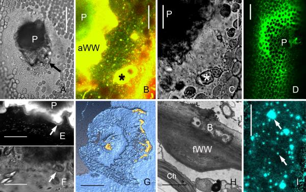
Wound walls induced by mechanical damage (puncturing), by bacteria and wound walls at alkaline regions of a cell exposed to high light intensity. (A-G) Puncture wounds in internodal cells of Chara corallina. (A) Bright field image of a healed wound. A fibrillar wound wall (arrow) is seen around the wound plug (P). (B and C) FM1-43 (green) and Lysotracker red-stained organelles are deposited near the wound plug (P) and around damaged chloroplasts (*) in early stages of wound healing (15 min after puncturing). C is the corresponding bright field image. (D) DiOC6-stained ER deposited at a wound 30 min after puncturing. (E and F) Wound plug (P) and FM1-43-fluorescent amorphous wound wall of a healed wound. A continuous FM1-43-fluorescent plasma membrane (arrow) is seen along the non-fluorescent inner, fibrillar wound wall 60 min after puncturing). F is the corresponding bright field image. (G) 3-D reconstruction of the inner surface of a healed puncture wound showing parts of the callose containing outer wound wall stained by aniline blue (yellow) and the calcofluor-labelled inner cellulosic wound wall (cyan), which is continuous with the normal cell wall. (H) Fibrillar wound wall (fWW) beneath bacteria (B) digesting the cell wall of an internodal cell of Nitella flexilis. Ch = chloroplast. (I) Wound walls (arrows) stained by Calcofluor at an alkaline region of a C. corallina internodal cell exposed to high light intensity (50 μmol m−2 s−1) for 10 days. Bars are 1 μm (H), 10 μm (B, C, E, F), 40 μm (A, D), 50 μm (I) and 75 μm (G)

