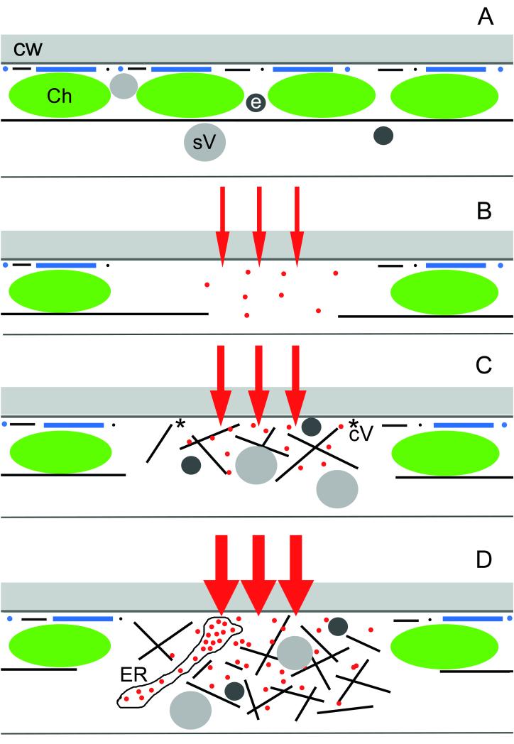
Fig. 6.
Schematic representation of early stages of wound responses in the cortex of characean internodal cells showing the major components involved, and the presumed Ca2+ influx. (A) In unwounded control cells and regions cortical actin filaments (black lines and dots) and microtubules (blue lines and dots) are present at the plasma membrane beneath the cell wall (CW). Continuous subcortical actin bundles are aligned along the stationary chloroplast (Ch) files. Secretory vesicles (sV) and putative endosomes (e) are present in the endoplasm and in the cortex. (B) Cortical window formation. Minor damage causes a small influx of external Ca2+ into the cytoplasm (red arrows and dots). This leads to the disassembly of the cortical cytoskeleton and of the subcortical actin bundles and to the detachment of chloroplasts. (C) Deposition of a fibrillar wound wall. Moderate Ca2+ influx causes accumulation of secretory vesicles and endosomes, which move along a newly formed actin meshwork. The presence of coated vesicles (cV) indicates that vesicle fusion with the plasma membrane is coupled with endocytosis of excess membrane. (D) Deposition of an amorphous wound wall. High Ca2+ influx causes not only accumulation of vesicles but also of Ca2+-loaded ER cisternae. Exocytosis of vesicles and ER takes place without endocytosis and excess membranes remain trapped within the amorphous wound wall.

