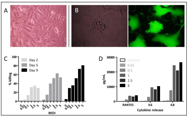
Figure 3.
Effects of MV on primary melanoma cells. A; Primary cells from freshly explanted melanoma B; Characteristic CPE 48 hours following treatment with MV-GFP by phase contrast (left) and fluorescence (right) microscopy. C; Live/Dead assay 2, 5 and 9 days after treatment of primary cells with MV. Data shown are representative of primary cells from three donors. D; ELISA of supernatant from primary cells treated with MV 72 hours previously.

