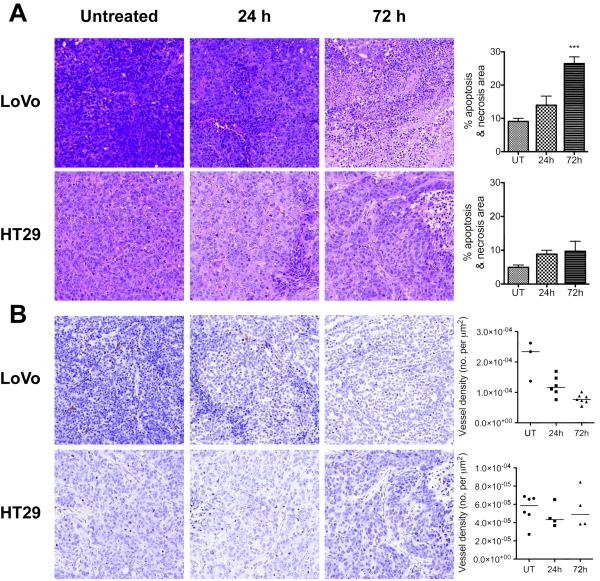
Figure 7.
Sections of tumors stained with hemotoxylin and eosin (A) and CD31 (B). Both tumors develop small abnormal or necrotic areas by 24h after the first Bevacizumab dose (A). In LoVo tumors, these expand significantly by 72h (p<0.001). Microvessel density (MVD) in the LoVo tumors is significantly higher than that of the HT29 tumors (B) before treatment (p<0.001) and the vessels are larger in size. After treatment, MVD decreases significantly at 24h (p=0.03) and 72h (p<0.001) in LoVo tumors, while remains unchanged in HT29 tumors.

