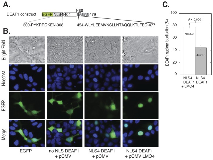
Figure 3. Nuclear localisation of EGFP-NLS4-DEAF1404–479 in the presence of LMO4.
A. DEAF1 construct in pEGFP-C2 that was used for transfection. It has an N-terminal EGFP tag followed by the altered DEAF1 NLS4 and DEAF1404–479. The NLS4 and NES protein sequences and DEAF1 numbering are shown. B. HEK293 cells grown on cover slips in 6 well plates were transfected with a total of 4 µg of DNA: control pEGFP (panel 1), 2 µg EGFP-DEAF1404–479+2 µg empty pCMV vector (panel 2), 2 µg EGFP-NLS4-DEAF1404–479+2 µg empty pCMV (panel 3) and EGFP-NLS4-DEAF1404–479+2 µg pCMV LMO4 (panel 4). After 24 h transfection, cells were fixed with paraformaldehyde and nuclei stained with Hoechst dye. Cells were imaged for EGFP fluorescence (green) and nuclear staining (blue) by fluorescence microscopy. C. Quantification of A. The two-dimensional areas of n = 8 fields of view were measured for % nuclear localisation of EGFP-NLS4-DEAF1404–479 in the presence and absence of LMO4. Difference is statistically significant as p<0.05.

