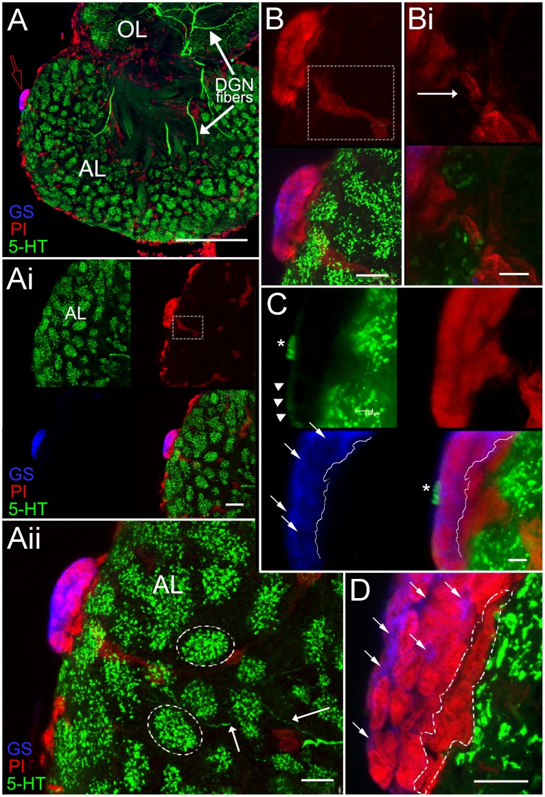
Figure 3. Stacked confocal scans of a 100 µm sagittal section in a P. clarkii brain show the neurogenic niche (A–D), stained for GS (blue), protruding from the ventral surface (arrow, red outline) of the accessory lobe (AL).
Serotonergic labeling (5-HT) in the olfactory lobe (OL) and AL (A–D, green) reveals fibers from the Dorsal Giant Neuron (DGN) innervating these lobes (A and Aii; white arrows). 5-HT labeling of individual glomeruli (dotted outlines in Aii) and DGN fibers (Aii, arrows) are clearly shown in this section of the AL. Magnifications of this section, including images deeper into the tissue (Ai, B, C) display the three separate laser channels representing the nuclear stain, propidium iodide (PI, red), serotonin (5-HT, green) and glutamine synthetase (GS, blue); PI and GS labeling together reveal the multi-layered aspect of the niche when the brain is sectioned sagittally (B, C, D). In C and D, white arrows indicate the GS-labeled cytoplasm (blue) of the niche cells, demonstrating that the deepest, most dorsal niche layer (outline in D) does not label for GS (dotted line in C, lower panels). The PI labeling cells also delineates the trail of cells, composing a blood vessel that extends from within the AL into the niche itself (Ai, white square; B,white square; Bi, arrow). Higher magnifications show a serotonin immunoreative “crown” of terminals within the outer, ventral layer of the niche (C, asterisk); a weakly immunoreactive serotonergic fiber(s) (arrowheads) is observed connecting to this serrated-like region. Scale bar: (A) 200 µm; (Ai, Aii, B) 20 µm; (D) 10 µm; (Bi, C) 5 µm.

