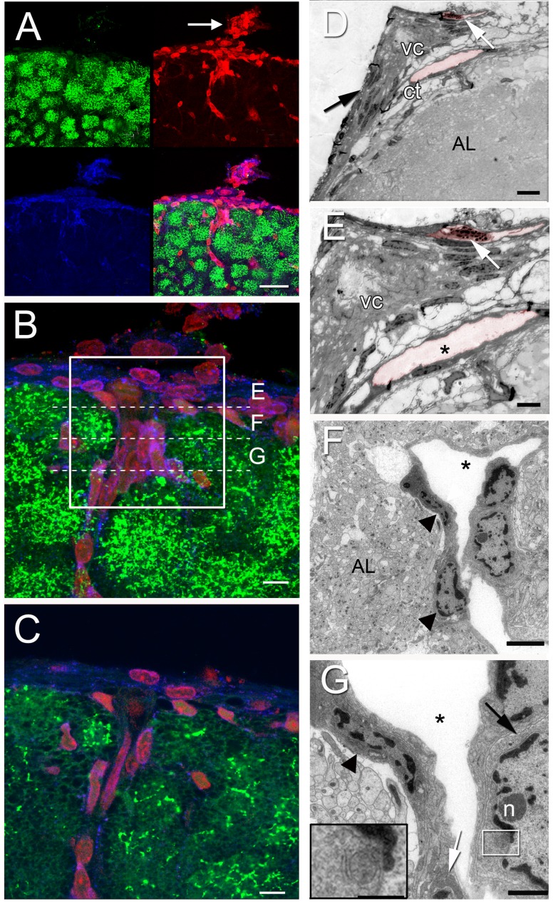
Figure 4. Confocal images of a sagittal niche section (A–C) with separate channels displayed in A (5-HT, green; GS, blue; PI, red; arrow pointing to the niche in A), reveal more dorsal aspects of the niche (B and C) and how cells are organized in regions just below the niche.
Corresponding semi-thin (E) and ultra-thin sections (F–G) through regions indicated in B reveal perivascular cells lining a blood vessel. (D, E) A semi-thin section in two magnifications. Note the vascular cavity (vc) and the lumen of two vessels, one below and one above the niche (colorized in pink). Within one of them a granular hemocyte is evident (white arrow). Observe the thin layer of connective tissue limiting the outermost part of the niche (black arrow, D), and in the innermost region, the loose connective tissue (ct) where there is a vessel. (F, G) Electron micrographs of a vessel dorsal to the niche in two magnifications, showing the perivascular cells (arrowheads) lining the vessel, with nuclei composed of heterochromatin apposed to the nuclear envelope, and euchromatin, which constitutes the major part of the nucleoplasm. Also, a nucleolus (n) is seen in one of the nuclei. (G) The cytoplasm of the perivascular cells (arrowhead) showing mitochondrial profiles (white arrow and insert [higher magnification of area limited by the white square]) and many endoplasmic reticulum cisternae (black arrow). Accessory lobe (AL in D and F). Scale bars: (A) 50 µm; (B and C) 6 µm; (D) 35 µm; (E) 20 µm; (F) 5 µm; (G) 2 µm; Insert in (G), 1 µm.

