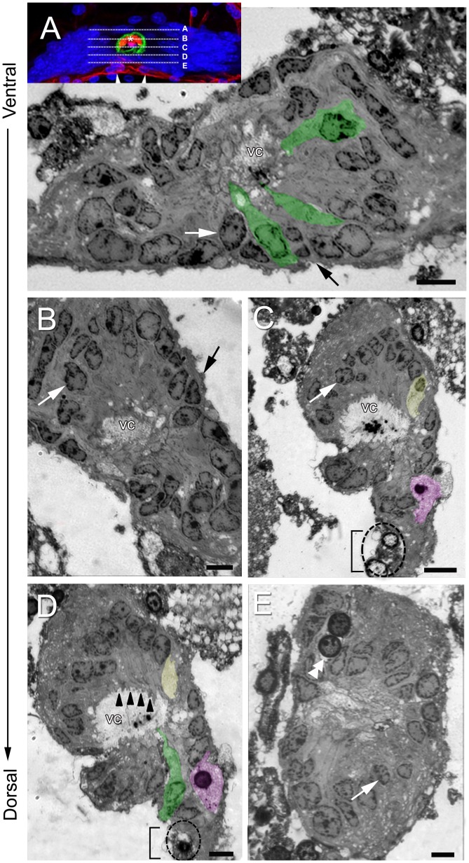
Figure 5. Semithin sections (ventral to dorsal sequence) of the crayfish niche stained with toluidine blue showing four types of cells, the outer edge of the niche and the central vascular cavity (vc).
Note that the images displayed in B–E are turned 90° relative to the image displayed in (A). (A, B) Type I cells (white arrows) are the most common, and have strongly stained cytoplasm and nuclei that are, in general, cuboidal or elongate. The cytoplasm of these cells lines the vascular cavity (examples are colorized green) and has fine extensions projecting into the lumen. The outer edge of the niche is covered with a thin layer of connective tissue (black arrow). The insert in (A) shows the position of corresponding semithin sections through the niche (A–E). (C, D) In more dorsal positions, in addition to Type I cells and their projections to the lumen (irregular cytoplasmic process – arrowheads in D), a Type II cell (colorized yellow) and Type III cells (one example is colorized purple) are identified. Type II cells have a nucleus with euchromatin and scarce heterochromatin, and a clear cytoplasm. The image in (D), which is 1.0 µm more dorsal than that in (C), shows only the cytoplasm of the Type II cell). Note two round cells (dotted circle) near to the emergence of the streams (bracket) with features that are distinct from the other cell types. These resemble Type IV cells (see below). Type III cells appear as amoeboid-shaped cells. Two type III cell nuclei are seen in the image (C), however only one appears in the image (D). Note the clear cytoplasm displayed by type III cells. In the center of the vascular cavity, there is intensely-stained material with a globular organization. The stream emerging from the niche is identified by the bracket. (E) Type IV cells (double arrowhead) have a spherical profile and are distant from the vascular cavity, near the emergence of the streams. This cell type frequently appears with paired nuclei and a clear cytoplasm. Type I cells are also seen (white arrow) in this image. Scale bars: (A–E) 12 µm.

