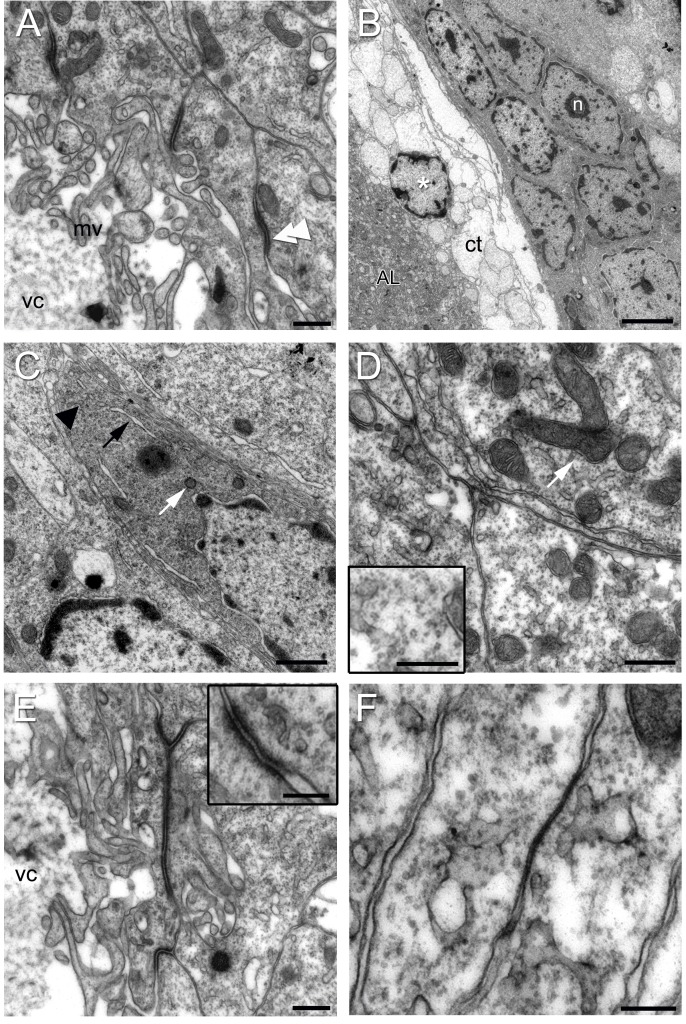
Figure 7. Electronmicrographs of Type I cells.
(A) Region of the niche near the vascular cavity (vc) showing Type I cells lining the edge of the cavity. Note the microvilli (mv) projecting into the cavity. Zonulae adherens join adjacent cells (double arrow head). (B) Type I niche cells near the emergence of the streams have elongated nuclei containing heterochromatin and euchromatin. A nucleolus (n) is apparent. Connective tissue (ct) is adjacent to the dorsal surface of the niche facing the accessory lobe (AL); a cell with features of a crustacean hemocyte (asterisk; hyaline cell, e.g., [56], [57]) is located within this tissue, although a blood vessel is not apparent. (C) Type I cells contain an abundance of mitochondrial profiles (white arrow), rough endoplasmic reticulum (black arrow), polysomes (arrowhead) and vesicles. (D) Cytoplasm of type I cells in high magnification: Observe the numerous mitochondria (arrow) and microtubules, seen in detail in the insert, lower left. (E) Another view of the apical surface of a Type I cell, showing a zonula adherens between adjacent cells, as well as microvilli projecting into the lumen of the vascular cavity (vc). The insert (upper right) shows a higher magnification of a zonula adherens. (F) Type I cells united by a septate junction. Scale bars: (A) 0.5 µm; (B) 5 µm; (C) 1 µm; (D, E) 0.5 µm; Inserts in (D) 0.1 µm and (E) 0.5 µm; (F) 0.2 µm.

