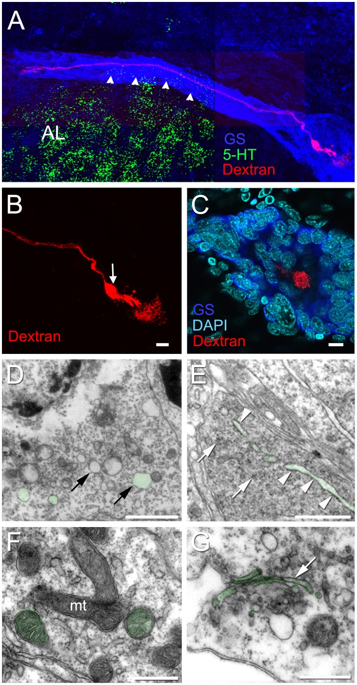
Figure 10. Alex Fluor® 568 hydrazide dye injection of Type I niche cells reveals their bipolar morphology.
Each Type I cell has a long process that stretches to either cluster 10 or cluster 9. (A) Montage showing the long process of a dye-injected cell, projecting towards cluster 10. Arrowheads point to the fascicle of Type I cell processes, labeled for GS, that form the migratory stream, and a short process that extends to the vascular cavity (B). Arrow in (B) points to the cell body, where the dye was injected. The injection of dye into Type I cells results in deposition of the dye in the vascular cavity (B and C). (C) A dye fill of a different Type I cell in which the niche is labeled for GS and stained with DAPI, shows the vascular cavity containing Alexa Fluor®, surrounded by the niche cells. (D–G) Additional ultrastructural details of the cytoplasm of Type I cells. (D) Vesicles (arrows). (E) Note rough endoplasmic reticulum (arrowheads) and some clumps of ribosomes (arrows). (F) Mitochondria (mt). (G) Golgi apparatus (arrow). Examples are colorized green. Scale bar: (B, C) 10 µm; (D) 2 µm; (E) 1 µm; (F) 0.5 µm. (G) 0.5 µm.

