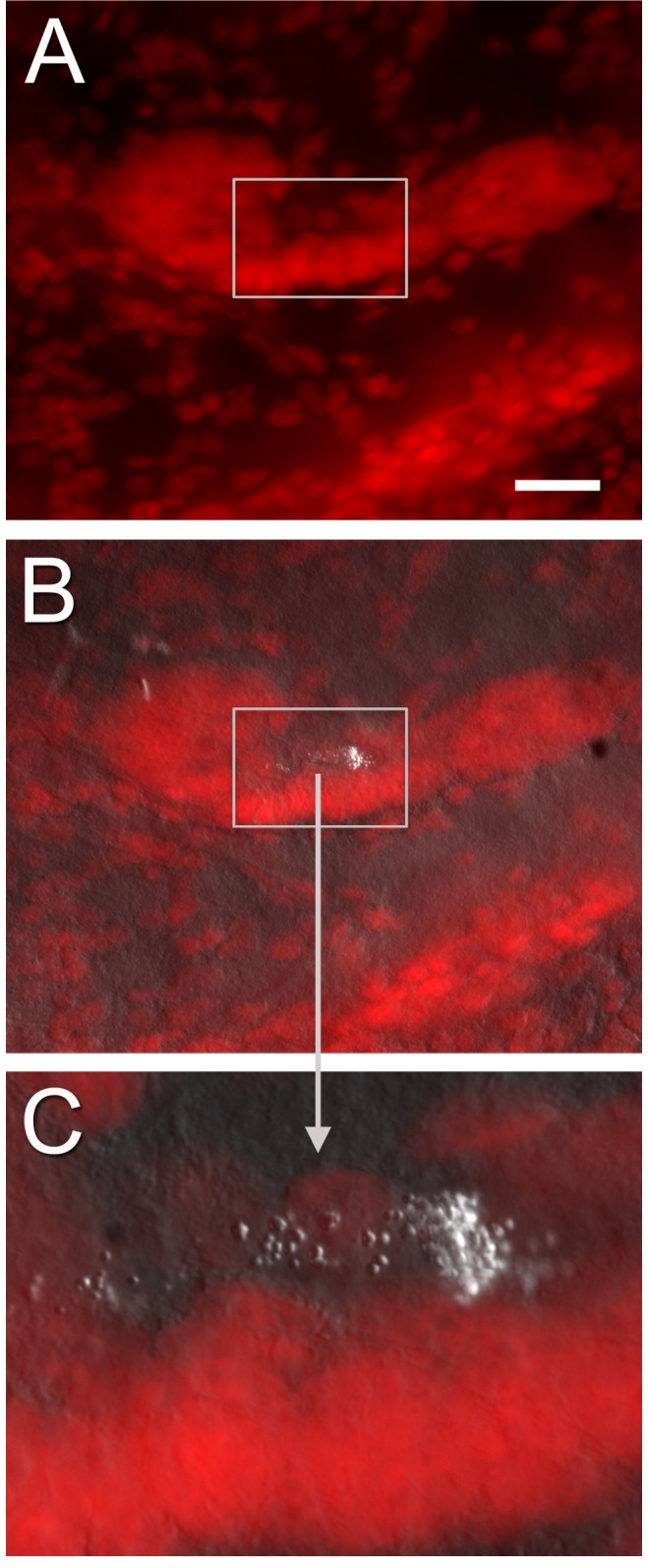
Figure 11. Combined confocal images (B, C) of PI-labeled cells in the niche (A) and Nomarski optics showing bubble-like material in the vascular cavity (higher magnification in C; rectangle in A and B).

These structures of unknown origin are also observed in EM sections (e.g., 9B–C, E–F) that show the material to be composed of electron dense particles. Scale bar: (A) 40 µm.
