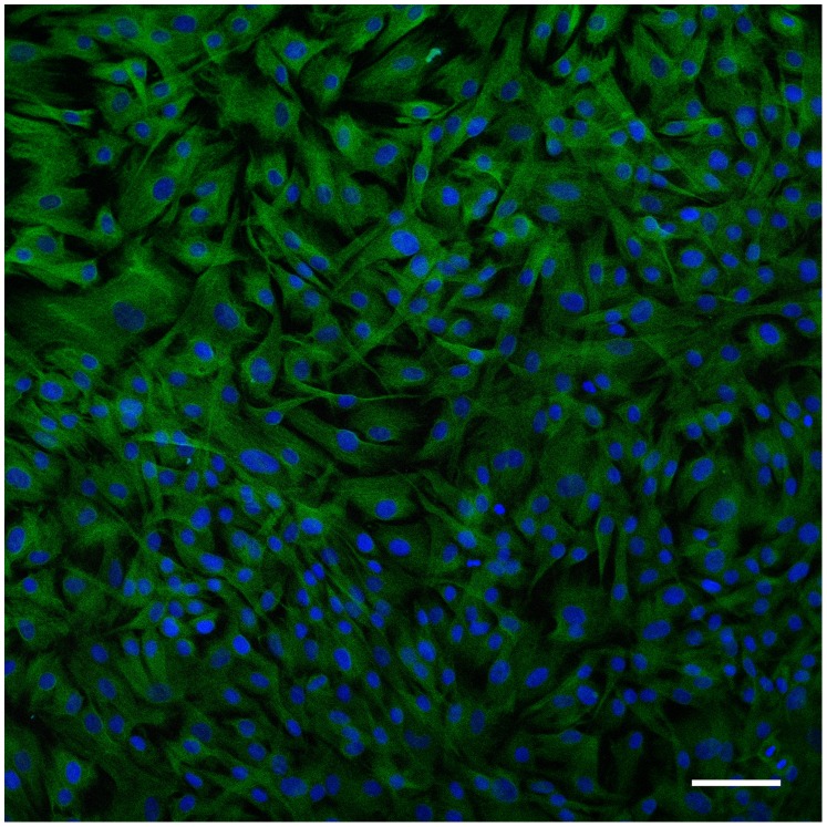
Figure 1. Confocal image of pterygium cells in culture stained with antibodies against vimentin (green), CD31 (white), and keratin 4 (red).
DAPI was used as a nuclear counterstain (blue). All cells were vimentin-positive and no immunoreactivity was found for markers of epithelial or endothelial lineages. Bar = 20 µm.

