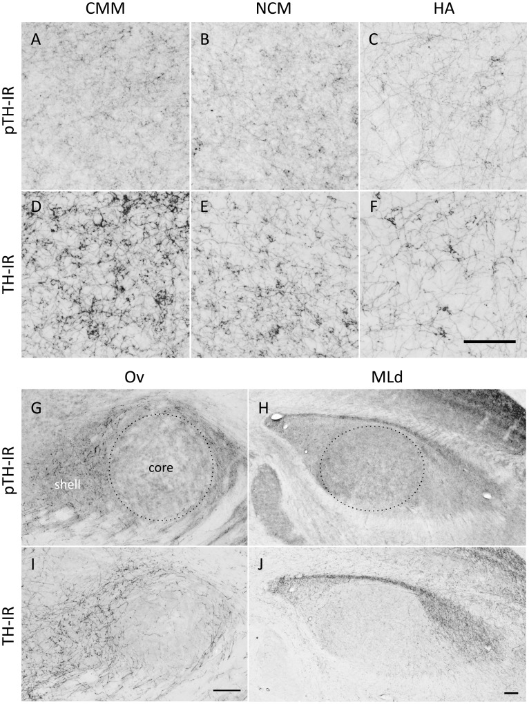
Figure 2. Examples of immunoreactive fibers labeled in this study.
Immunoreactivity (IR) for phosphorylated tyrosine hydroxylase (pTH; A, B, C, G, H) or tyrosine hydroxylase (TH; D, E, F, I, J) is shown in the caudomedial mesopallium (CMM; A, D), caudomedial nidopallium (NCM; B, E), apical hyperpallium (HA; C, F), n. Ovoidalis (Ov; G, I) and the dorsal lateral mesencephalic nucleus (MLd; H, J) in birds that heard 15 min of song. Dotted lines encircle the areas sampled in the Ov core and MLd. Rostral is to the right. Scale bars, 100 µm.

