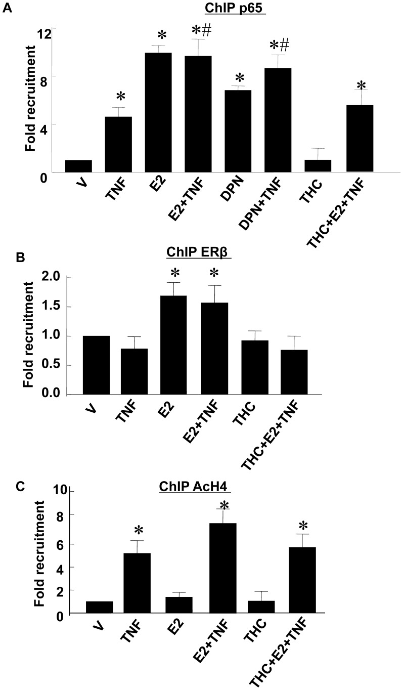
Figure 5. ChIP assays of the binding of NFκB p65 (A), ERβ (B) and AcH4 (C) to the IκBα promoter.
Cells were pretreated with/without E2 (10−7 M) or DPN (10−7 M) for 24 hrs and then stimulated with TNF-α (1 ng/mL) for 1 hr. THC (10−6 M) was given to cells at 1 h before E2 treatment in some experiments. ChIP samples were prepared as described in the text and analyzed using antibodies specific for p65, ERβ or AcH4. The immunoprecipitated DNA fragments and input DNA were analyzed by real-time PCR. The y axis shows values were normalized to input DNA with values for vehicle treatment defined as 1. The numbers represent the mean±SEM from three experiments repeated in duplicate. *p<0.05 vs. Vehicle-treated RASMCs; #p<0.05 vs. TNF-α-treated RASMCs.

