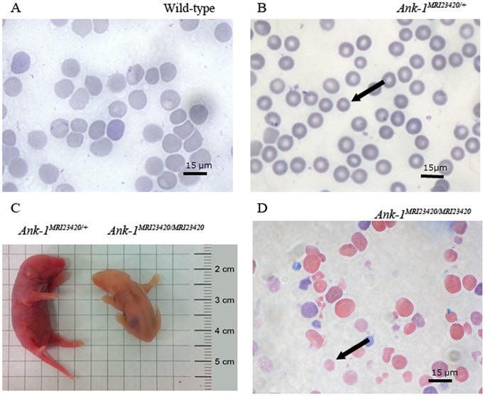
Figure 3. Identification of Ank-1MRI23420/+ and Ank-1MRI23420/23420 mice.
Giemsa stained peripheral blood smears from (A) wt, (B) Ank-1MRI23420/+, and (D) Ank-1MRI23420/MRI23420 mice. Black arrows indicate microcytic RBC in Ank-1MRI23420/+ and spherocytes in Ank-1MRI23420/23420. (C) Jaundiced postnatal day 1 of Ank-1MRI23420/MRI23420 pup and an Ank-1MRI23420/+ control littermate.

