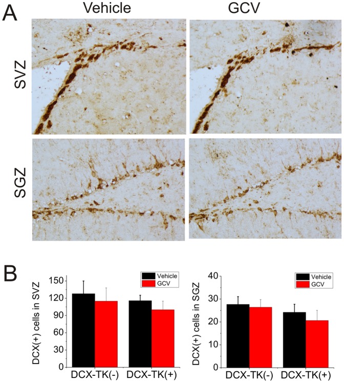
Figure 4. DCX-immunopositive cells in SVZ and dentate SGZ of vehicle- and GCV-treated, wild-type and DCX-TK transgenic mice, 12 weeks after MCAO.
(A) Representative images of DCX-immunoreactive cells in SVZ (top) and dentate SGZ (bottom) from vehicle (left)- and GCV (right)-treated DCX-TK(+) transgenic mice. (B) Quantification of DCX-immunoreactive cells in SVZ (left panel) and dentate SGZ (right panel) from GCV (red bars)- and vehicle (black bars)-treated DCX-TK(+) and DCX-TK(-) mice. There were no significant differences between vehicle- and GCV-treated groups.

