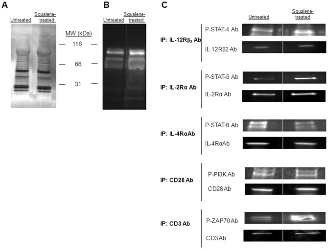
Figure 3. Tyrosine phosphorylation patterns of major signaling modules of T helper differentiation upon enrichment of membrane cholesterol.
(A) SDS-PAGE silver stain of pooled, negatively-sorted CD4+ splenic T-cell lysates from F1 mice (n = 4/group) untreated (left lane) or squalene treated (single dose of 180 µg, right lane) 3 days post-injection shows no detectable quantitative alterations in the protein bands between the two groups of mice. (B) Tyrosine phosphorylation patterns of the same samples in panel A were blotted with anti-phosphorylated tyrosine Ab-HRP conjugate. Of note, the amount of 55–100 kDa phosphorylated protein bands is increased in mice treated with squalene. (C) Immunoprecipitation of pooled splenic CD4+ T-cell lysates from F1 mice treated or not with squalene (180 µg/mouse, n = 4/group) 3 days post-injection was carried out for IL-12Rβ2, IL-2Rα, IL-4Rα, CD28, or CD3 receptors using specific Abs, and probed with specific anti-phospho Abs for STAT4, STAT5, STAT6, PI3K, and ZAP-70 kinases. Only the phosphorylated STAT-4, STAT-5, and ZAP-70 in squalene treated mice were significantly enhanced. Shown is one of two representative experiments.

