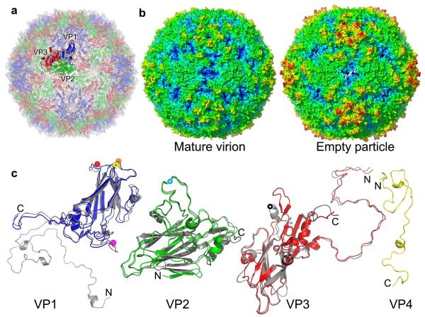
Figure 1.
Overall structures. (a), Cartoon of the mature EV71 virion, looking down an icosahedral 2-fold axis; VP1, VP2, VP3 and VP4 are drawn in blue, green, red and yellow, respectively. A single icosahedral protomer is drawn more brightly. (b), Radius colored surface representation of EV71 mature and empty particles. The surfaces for both are colored from blue to red according to their distances from the particle center. (c), Structures of the EV71 capsid proteins. VP0-3 (we treat VP0 and VP2 interchangeably) are shown in a similar orientation to the brighter protomer in (a); proteins from the expanded empty particle are coloured as in (a), whereas the corresponding chains from the mature virion are grey. VP4 of the mature virion is shown in yellow. The proteins have been superimposed as rigid bodies (216, 227, 220 Cαs superpose with rms deviations of 1.6, 0.9 and 1.3Å for VP1, VP0(2) and VP3 respectively). Residues 1-297 of VP1, 10-254 of VP2, 1-242 of VP3 and 12-69 of VP4 have well defined electron density in the mature virion, while residues 73-210 and 219-297 of VP1, 82-319 of VP0 and 1-238 of VP3 in the empty particle are modeled. Major surface exposed loops are marked with coloured washers: VP1 BC (yellow), VP1 DE (orange), VP1 HI (red), VP1 GH (magenta), VP2 EF (light blue), VP3 GH (black).

