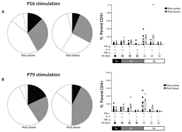
Figure 4. Multifunctionality of P16 and P79 specific CD4+ T cells.
Mice were immunized with the same regimen as described in Figure 1. Two weeks post-prime with AdHu5-PcAMA1 (day 14) and two weeks post-boost with MVA-PcAMA1 (day 70), splenocytes were re-stimulated with the P16 and P79 peptide or no peptide (Unstim) for 5h. Cells were surface stained for CD4 and intracellularly stained for IFN-γ, TNFα and IL-2, and assessed by flow cytometry to determine the % of responding cells per total CD4+ T cell subset. Toal responses of each cytokine to P16 (A) and P79 (B) are shown at each time-point. The multifunctionality or “quality” of CD4+ T cells was analyzed using the SPICE software.

