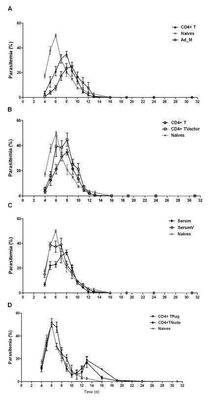
Figure 8. Adoptive transfer of CD4+ T cells and serum from vaccinated mice to normal and immunocompromised mice.
BALB/c mice were immunized as before with AdHu5-MVA PcAMA1. Two weeks after the MVA boost, spleens were harvested, pooled and the CD4+ T cells were isolated as described in Methods. 9 × 10⁷ CD4+ T cells from vaccinated mice were injected i.v. into naïve wild-type mice (CD4+ T), RAG1/2−/− mice (CD4+ TRag), or BALB/c athymic nude (nu/nu) mice (CD4+ TNude). CD4+ T cells from vector control immunized mice were also isolated and transferred into naïve wild-type mice (CD4+ TVector) in the same manner. Serum was also harvested from each vaccinated and vector control mouse, pooled, and then transferred into two groups of naïve wild-type mice (labelled as Serum and SerumV respectively). 500μl of serum was injected i.p. on days -1 and 0 (total 1ml into each mouse), with respect to challenge on day 0. A group of naïve wild-type mice and vaccinated (Ad_M) control mice were also included. n=5 mice/group except for the serum transfer groups where n=4/group. All mice were then challenged with 106 PccAS pRBC and montiored as previously. In the CD4+ TRag group, 3 mice died (one on each of days 8, 9 and 11 denoted by +) and in the CD4+ TNude group one mouse died (day 10 denoted by ++). Panels (A-D) show comparisons between the various groups and the mean group % parasitemia ± sem over time.

