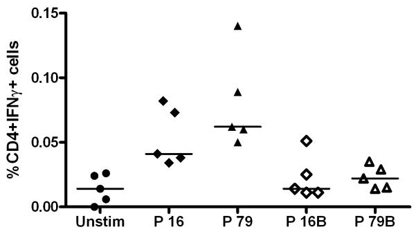
Figure 9. T cell response to heterologous AMA1 peptide sequences from other P. chaubaudi strains.
BALB/c mice (n=5) were primed with 1 × 10¹⁰ vp AdHu5-PcAMA1 and boosted 8 weeks later with 1 × 10⁷ pfu MVA-PcAMA1. Two weeks post-boost (day 70), splenocytes were isolated and re-stimulated with the peptides P16 and P79 (as described previously), or with P16B and P79B (heterologous peptides to P16 and P79, respectively), or no peptide (Unstim). Cells were surface stained for CD4 and intracellularly stained for IFN-γ, and then assessed by flow cytometry to determine % of responding cells per total CD4+ T cell subset. The figure shows the individual and median CD4+ IFN-γ+ responses.

