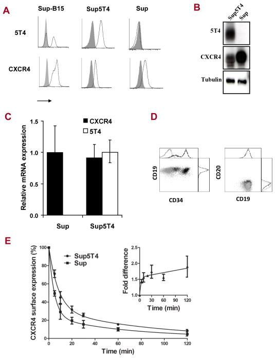
Figure 2. Characterization of Sup-B15 5T4+ (Sup5T4) and 5T4− (Sup) sublines.
Analysis of CXCR4 and 5T4 expression by (A) Flow cytometry, (B) Western blot (C) qPCR. (D) Flow cytometric phenotyping for CD19, CD20 and CD34 (Sup=light grey; Sup5T4=black). (E) Average results from three independent experiments showing differential CXCL12 modulation (30ng) of cell surface CXCR4 expression (fluorescence index determined by flow cytometry) on Sup5T4 compared to Sup cells; * is p <0.05. After treatment with 30ng of CXCL12, the half-life of CXCR4 detected by flow cytometry is 10.6 and 5.8min for Sup5T4 and Sup respectively; a 1.8 fold difference in rate of CXCR4 modulation. (Inset) Fold difference in CXCR4 expression by Sup5T4/Sup with time of modulation by CXCL12. Similar results were seen at 100ng CXCL12 treatment.

