
Figure 2.
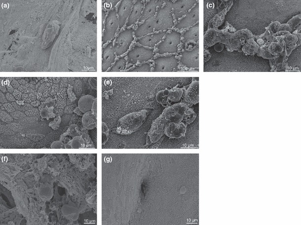
Comparison of colonisation of the colonic surface by Entamoeba histolytica and Entamoeba dispar. Panels show breakdown of mucus by E. histolytica after 0 h (a) and 2 h (b). Enlargement of region shows aggregates of trophozoites and recruited human cells (c). After 4 h, trophozoites begin to damage (d) and to penetrate epithelia (e). Conversely, after 4 h, E. dispar binds to, but does not degrade, the mucus barrier (f) and, as shown by manually removing the mucus layer, does not recruit immune cells to the epithelial surface (g). [Reprinted, with permission, from (44)].