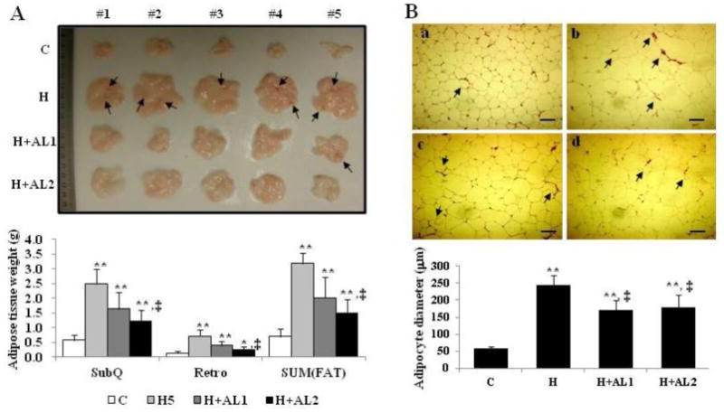
Fig. 3.

Images of the isolated subcutaneous adipose tissue (A) and adipocytes (B) of mice. (A) Isolated whole subcutaneous adipose tissues (top) and their measured weights (bottom) were analyzed. (B) Cryosectioned adipocytes (top) and their diameters (bottom) were measured. Data are presented as mean ± S.D. (n = 5). Scale bar = 200 μm. * and **indicate that the differences among the control (C-mice) and experimental mice (H, H+AL1, or H+AL2 mice) were statistically significant at p < 0.05 (*) or p < 0.01 (**). ‡ denotes the significant differences among H and AL treated groups (i.e. H vs. H+AL1 or H+AL2 mice group, p < 0.01).
