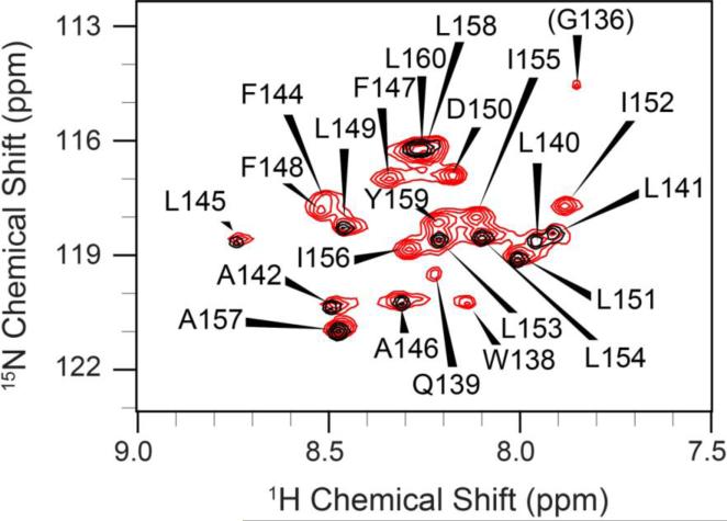
Figure 4.
2D 1H, 15N-HSQC spectra(excluding most the Gly region) of the TMD-5 peptides solubilized in bicelles. Red and black contours correspond to the spectrum of uniformly 13C, 15N enriched TMD-5 and selectively 15N-GLA labeled TMD-5, respectively. The spectrum shows nice chemical shift dispersion with well-defined peaks and line shapes, indicative of a folded, homogenous protein tertiary structure. Residues are numbered according their position in the native LMP-1 sequence.

