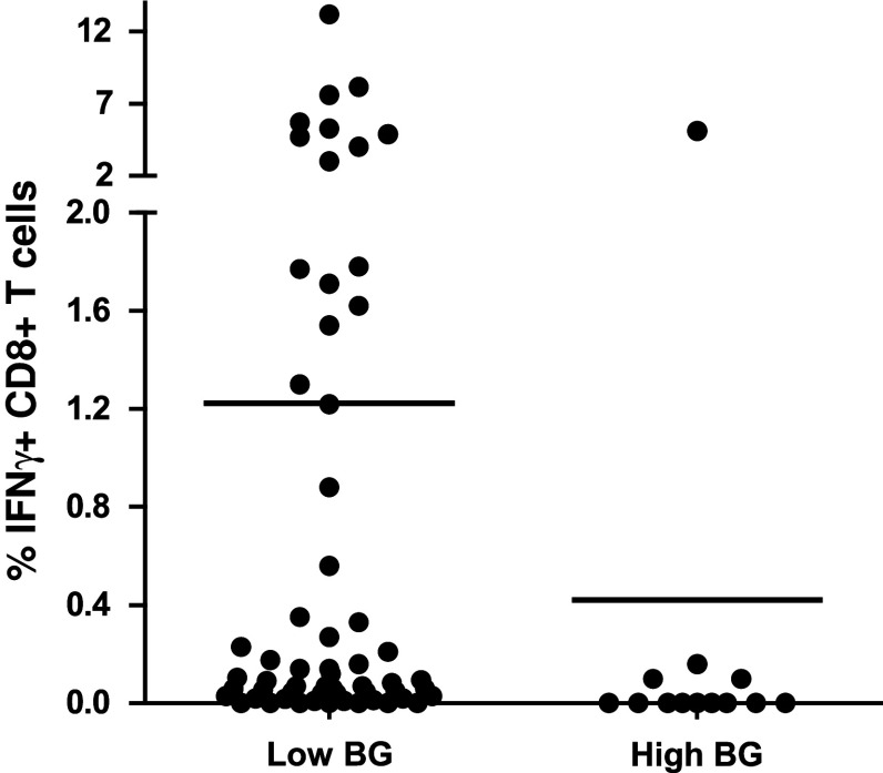
Fig. 2.
High background staining decreased ability to detect responses in phase 1. The detection (i.e., frequency of IFNγ-producing specific CD8+ T cells after FLU or CMV peptide stimulation) is shown only for the positive donor–antigen combinations versus a low or high background in the corresponding negative control sample. The background was classified as low (n = 59) or high (n = 13) based on the average background value (0.118 %) measured in all negative control samples (n = 72) accumulated from all participants

