Figure 5.
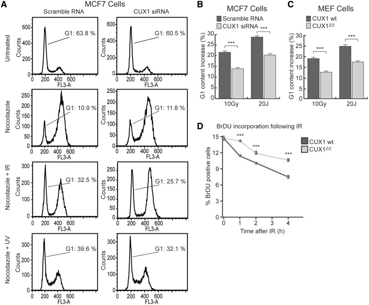
Effect of CUX1 knockdown on G1/S and S phase arrest following damage. (A) MCF7 cells were transfected with CUX1-specific siRNA and exposed to either 1 µm Nocodazole, Nocodazole + 10 Gy of IR or Nocodazole + 20 Js of UV. Cells were fixed with ethanol 24 h after exposure, stained with Propidium Iodine (PI) and analyzed for cell cycle distribution by flow cytometry. (B) A histogram of the increase in G1 content after IR and UV in MCF7 cells. *P < 0.05, **P < 0.01, ***P < 0.001; Student's t-test. (C) A histogram of the increase in G1 content after IR and UV in Cux1Z/Z and Cux1 wt MEF cells treated and analyzed as in A. (D) Cux1Z/Z and Cux1 wild-type MEF cells were exposed to 10 Gy IR. 1 to 4 hours post exposure, the cells were labeled with BrdU for 1 h before fixation with 4% Paraformaldehyde. BrdU incorporation was measured by flow cytometry.

