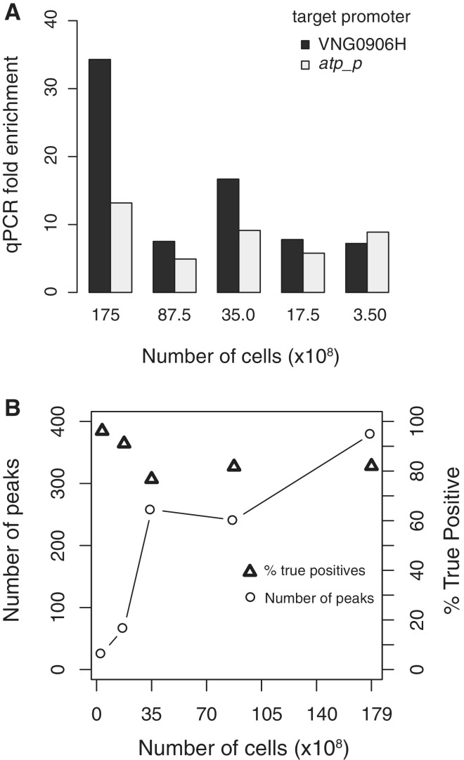
Figure 6.
TfbD ChIP with decreasing numbers of cells. (A) ChIP-qPCR determined fold enrichment at two test promoters (VNG906H, dark bars and atp_p light bars) decreases with lower numbers of cells; however, strong (>5 x) enrichment is still observed with 3.5 × 10⁸ cells. (B) For decreasing cell volume ChIP-seq experiments, fewer peaks could be identified (number of peaks identified, squares and lines), resulting in a significant loss in sensitivity. However, the percentage of identified peaks that were true positives stayed high (% true positives, triangles). True positives were defined as binding sites that were shared with at least one of the optimized 1.75 × 10¹⁰ ChIP TfbD biological replicates.

