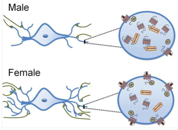
Figure 2.
This schematic illustrates sex differences in the LC-arousal system. The images on the left depict LC neurons with dendrites (blue) receiving afferent inputs (in green). Because the LC dendritic tree is longer and more complex in females, they are more likely to receive projections from afferent structures that terminate in the peri-LC. The images on the right depict magnified views of LC dendrites in order to illustrate sex differences in the CRF1 receptor coupling and trafficking. In males, CRF1 receptors (red) associate with β-arrestin2 following ligand binding and become localized in the cytoplasm. CRF1 receptors of females are highly coupled to Gs and following ligand binding they are predominantly found on the plasma membrane.

